TOTAL EAR CANAL ABLATION " TECA "

Total Ear Canal Ablation (TECA) is a salvage surgical procedure performed to remove the entire external ear canal, usually indicated for end-stage otitis externa, neoplasia, or irreversible ear canal disease

In cats, polypoid or neoplastic masses within the ear canal or middle ear can cause chronic infection, pain, and neurological signs, necessitating surgical management

History

Clinical Signs

Cytology

Treatment

Histopathology

A 10-year-old domestic short-haired cat was presented with:

Chronic ear discharge, foul odor, and persistent head shaking

Pain and swelling around the affected ear.

Occasional scratching and vocalization due to discomfort.

The condition had been unresponsive to medical therapy (topical and systemic antibiotics, anti-inflammatory drugs).

The external ear canal was stenotic and filled with purulent discharge

A polypoid mass was visualized obstructing the vertical canal

Palpation elicited marked pain

No neurological deficits were initially observed.


Cytology and culture of the discharge revealed mixed bacterial infection.
Fine-needle aspiration (FNA) of the mass suggested chronic inflammatory polypoid tissue, with no definitive signs of malignancy


Total Ear Canal Ablation was performed: complete removal of the vertical and horizontal ear canal

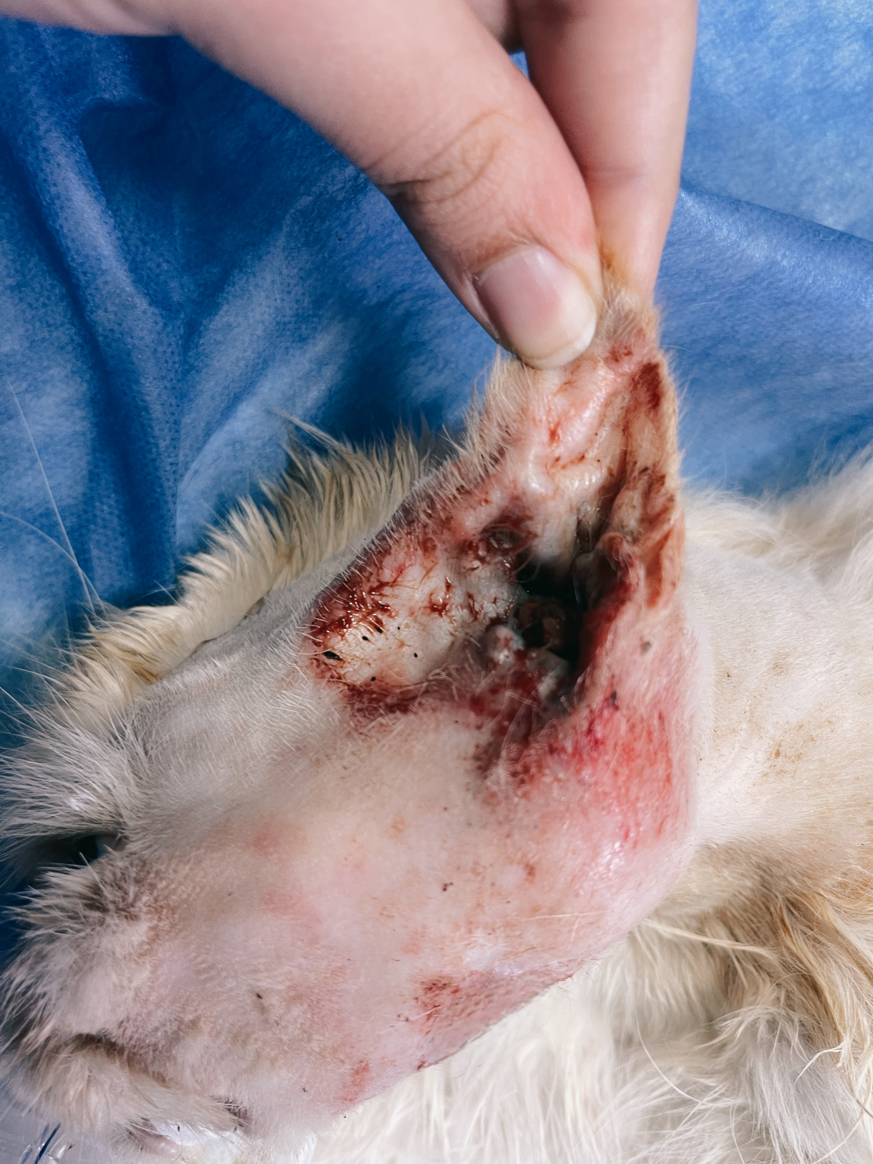
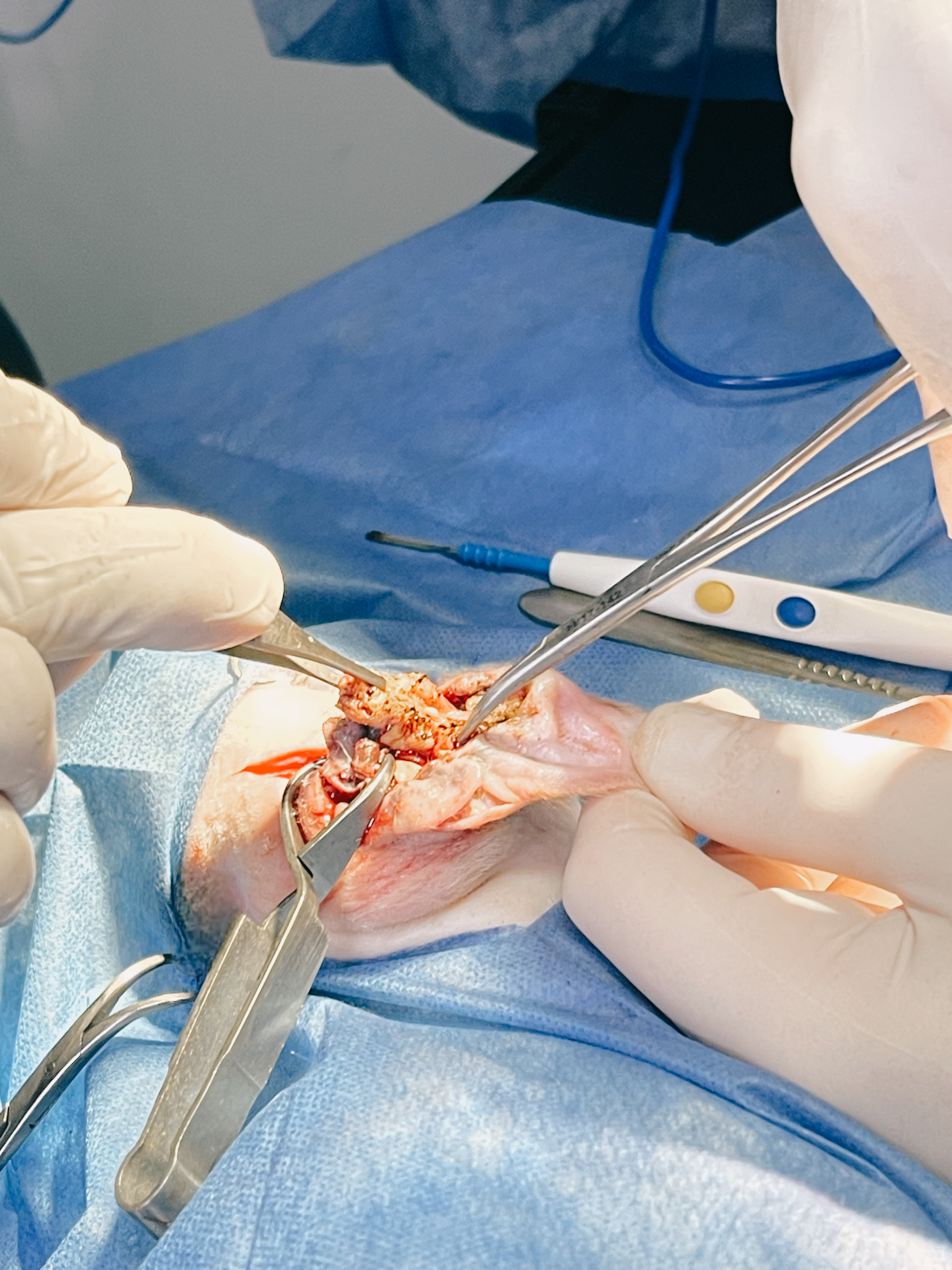
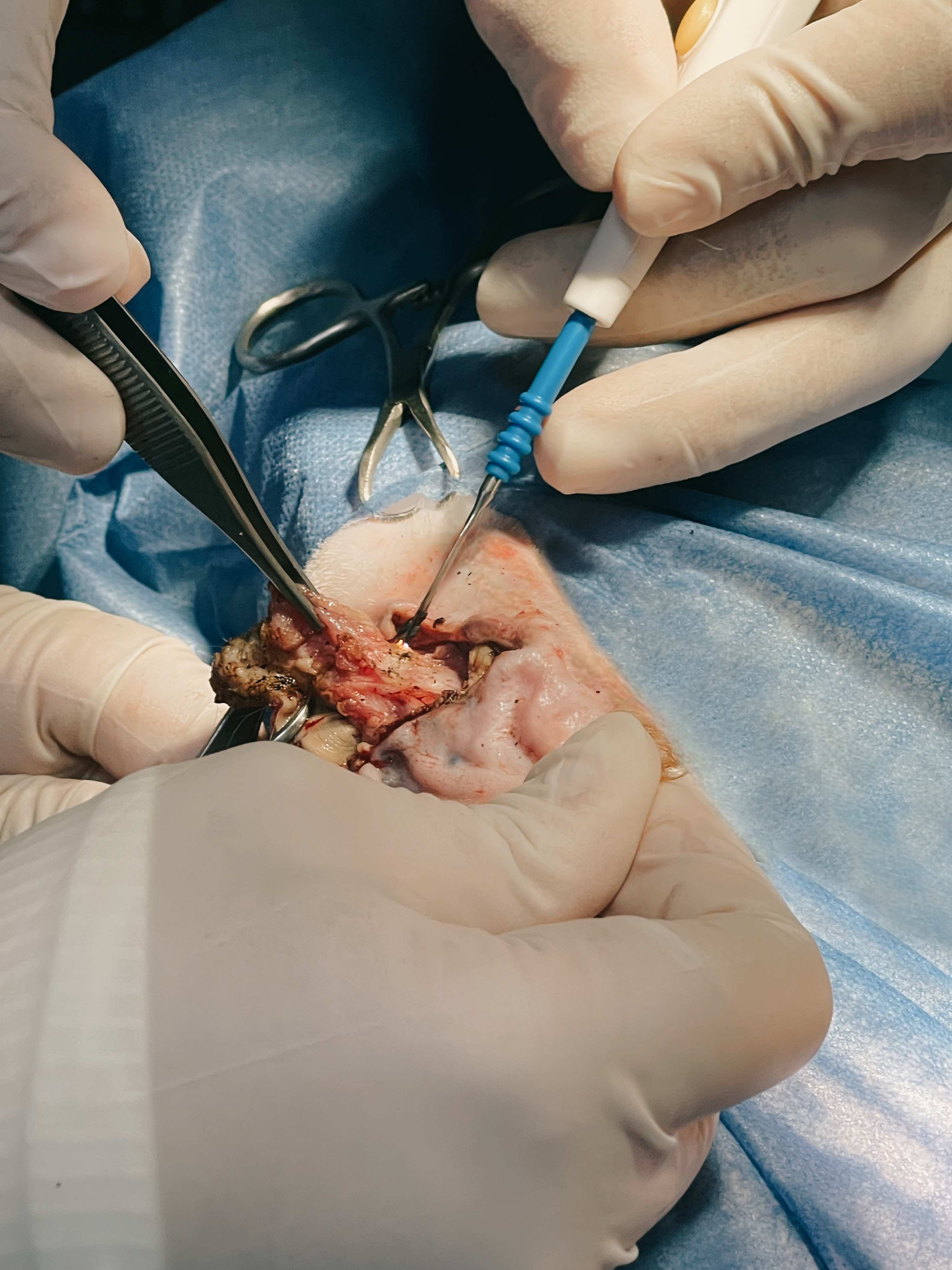
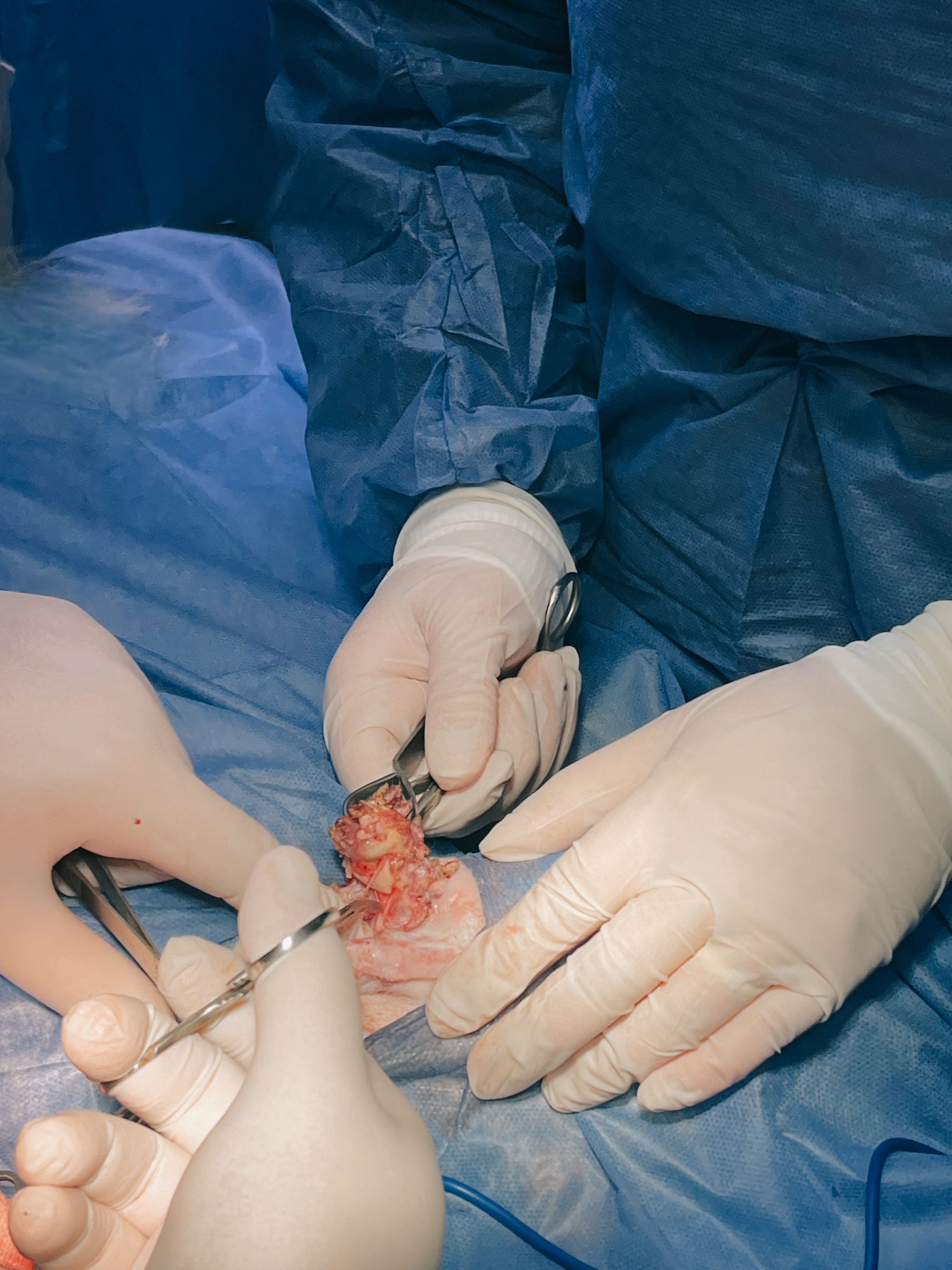
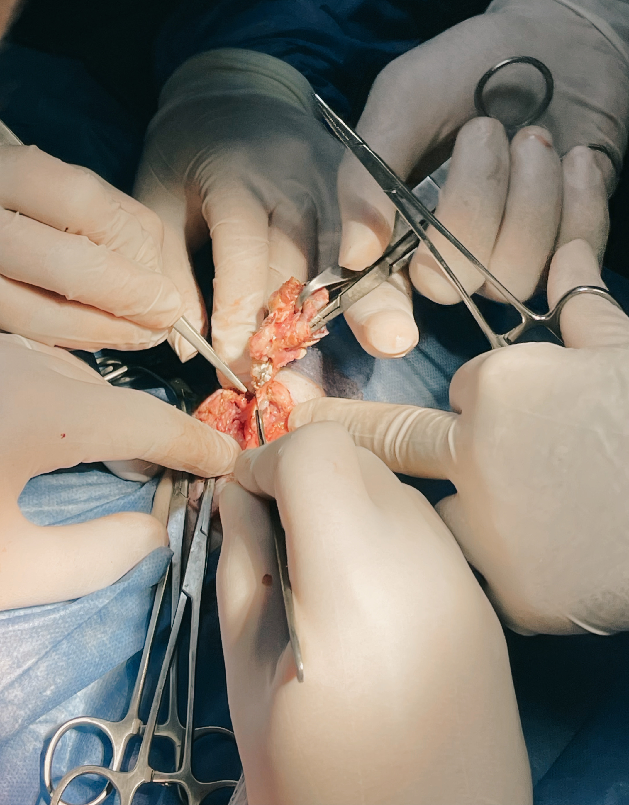
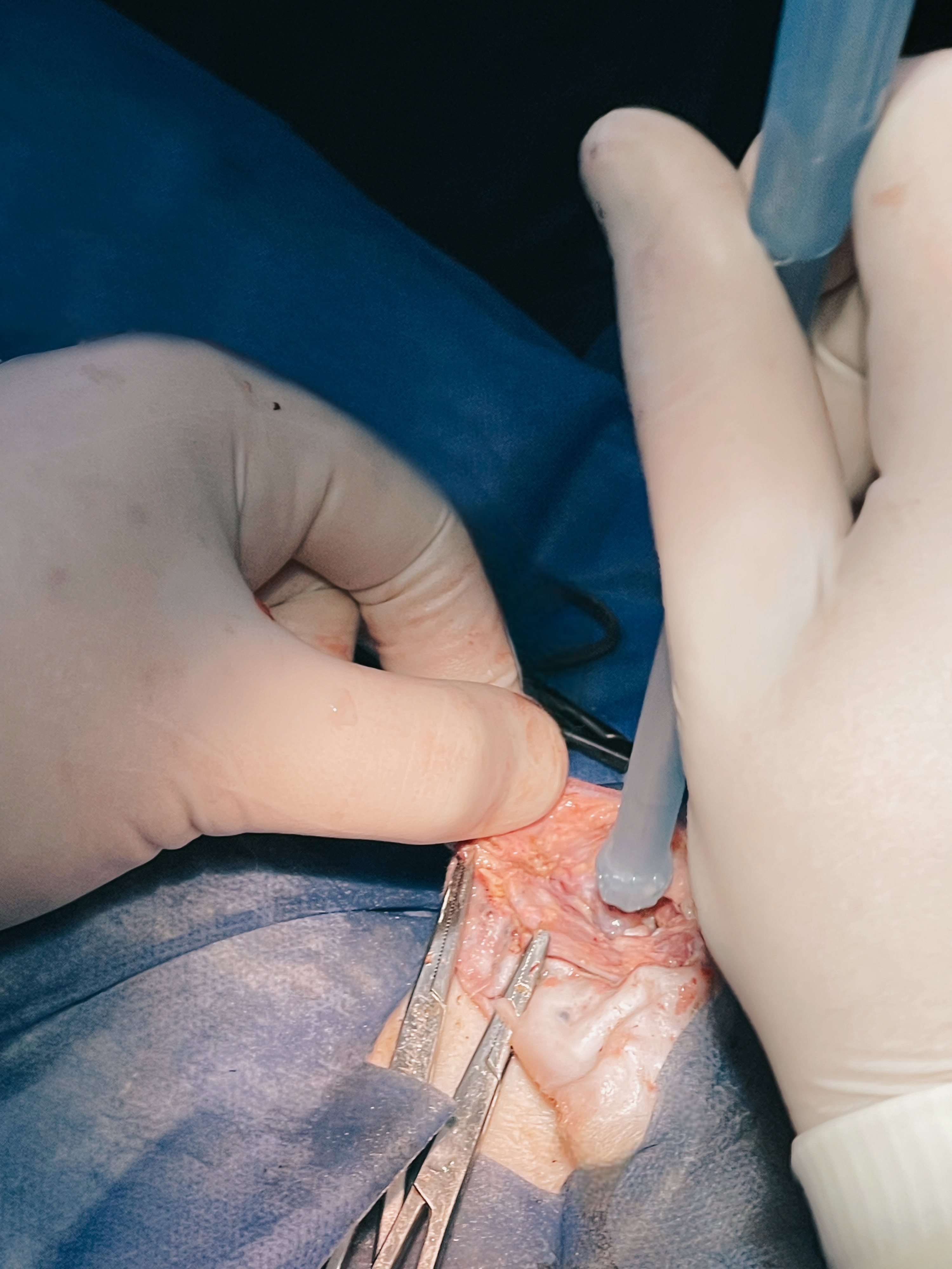
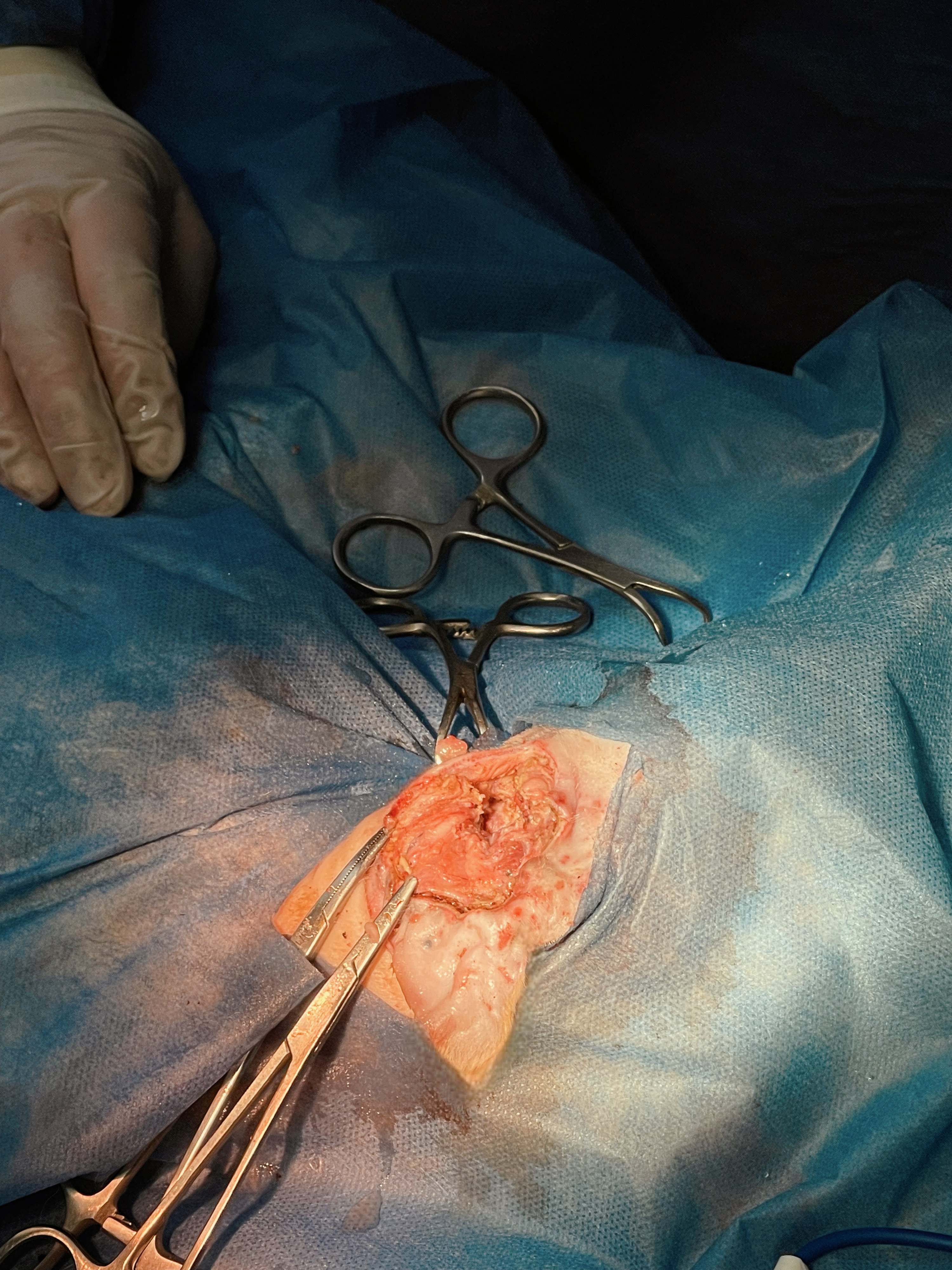
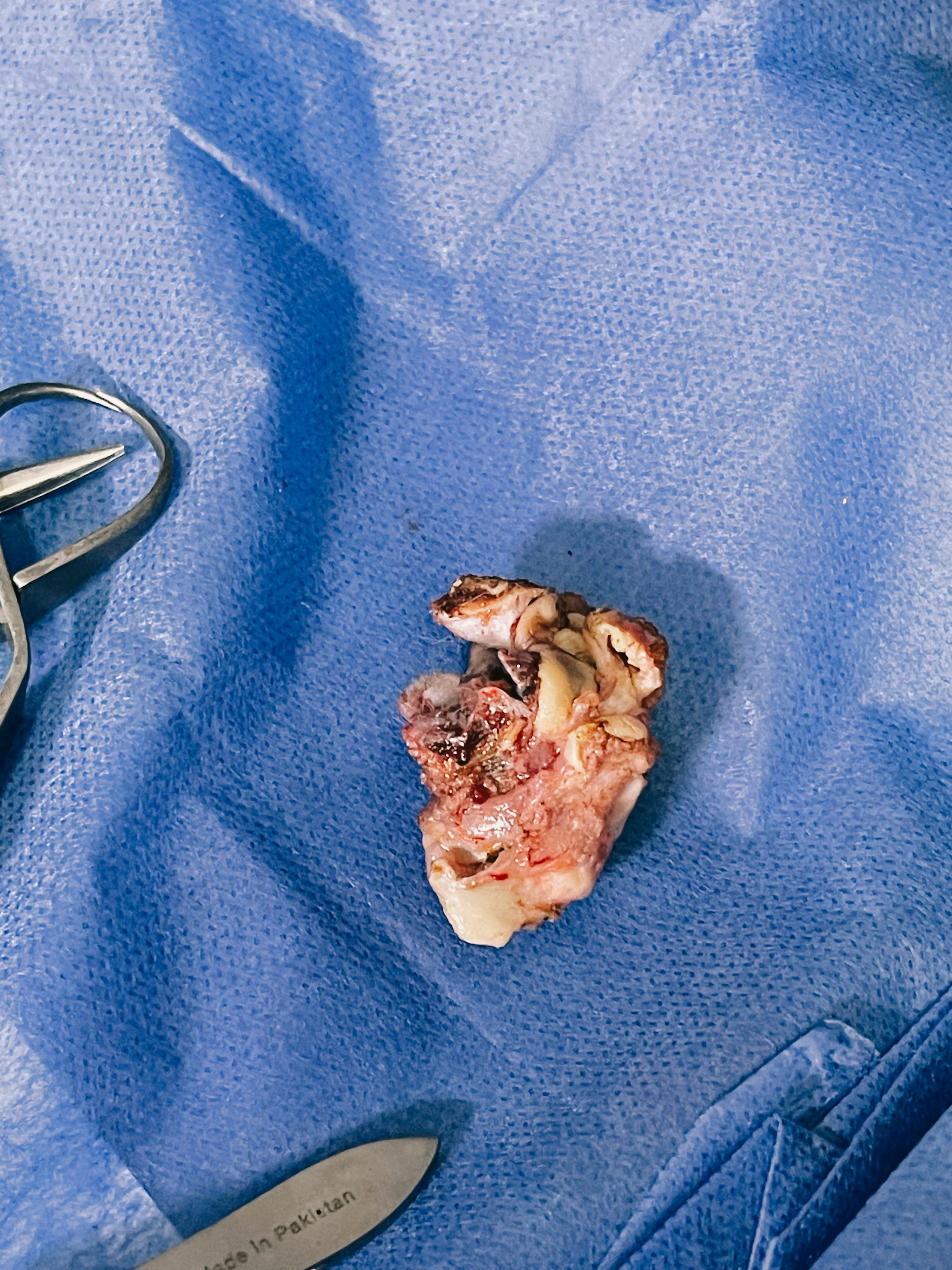
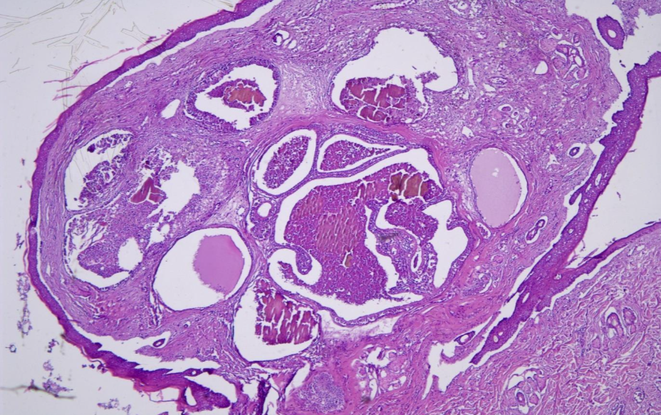
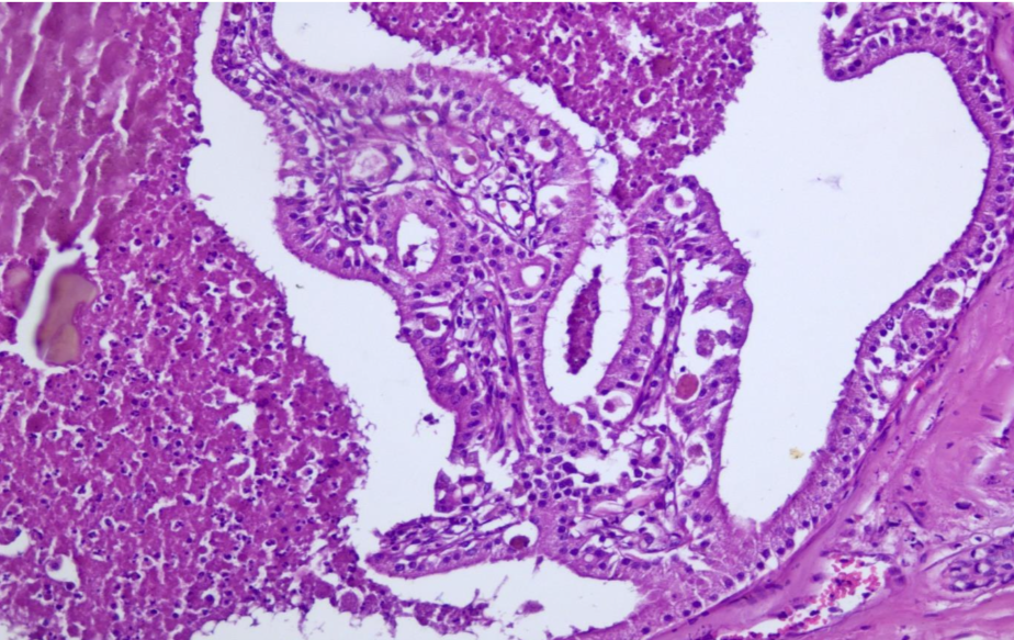
The deep dermis was expanded by an unencapsulated, well-circumscribed, multilobular neoplasm composed of two populations of cells (Fig. 1). The first population was composed of cuboidal to columnar cells forming varisized islands and well-differentiated tubules supported by a dense fibrovascular stroma. Neoplastic cells had indistinct cell borders, a small to moderate amount of granular eosinophilic cytoplasm (Fig. 2).

Within tubules, neoplastic cells frequently form few papillary projections into tubule lumina. Tubules contained necrotic debris and fragmented neutrophils. Ceruminous gland proliferation has areas of ectasia and hyperplasia as well as regional hyperplasia of sebaceous glands and pleocellular otitis externa.

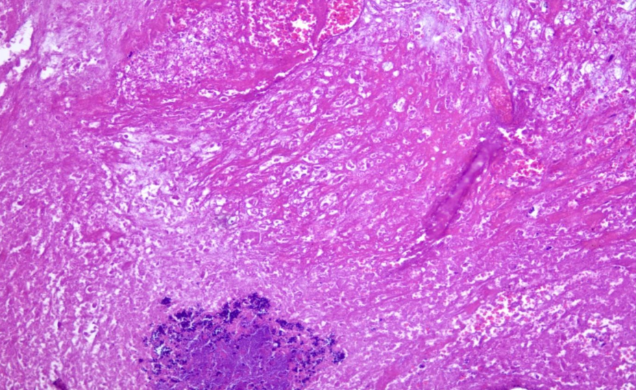
Some examined sections showed ulceration in the epidermal layer with extensive dermatitis that characterized by inflammatory cells infiltration (Fig. 3). Minimal pleomorphism was occurred and mitotic figures were few to absent. Inflammatory cells were infiltrating the dermis, which showing varying numbers of lymphocytes and neutrophils.

Conclusion: Ceruminous gland adenoma

.

.

.

.

.

.

.

.

.

.

.

.

Polyp excised

Histopathology

Histopathology

Histopathology

Explore Our New uploads

RECENT SURGICAL PROCEDURES

© All Rights Reserved