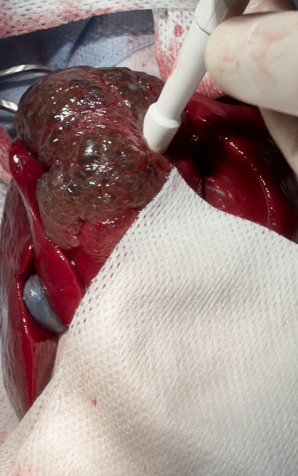
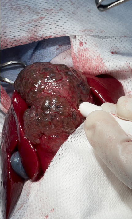
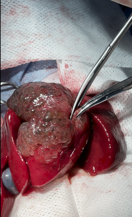
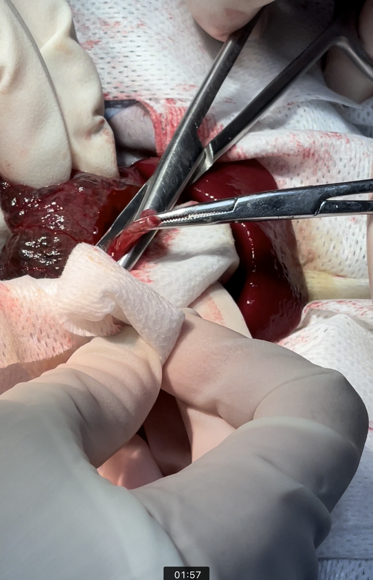
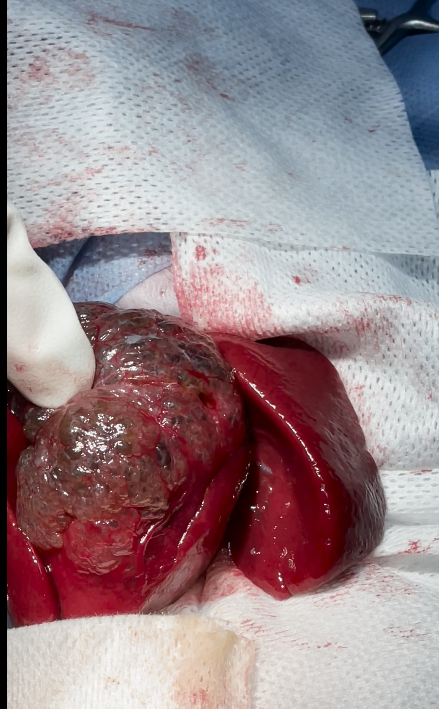
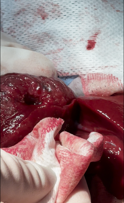
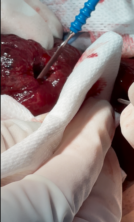
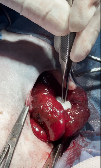
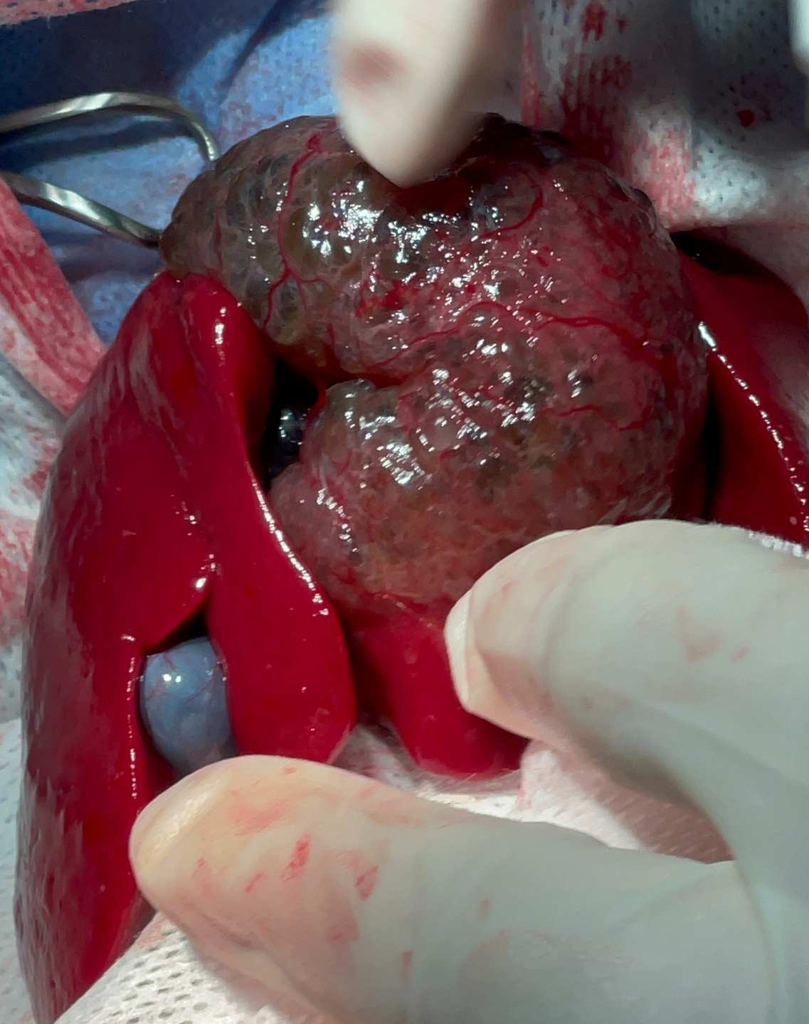
Hepatobiliary Cystadenoma

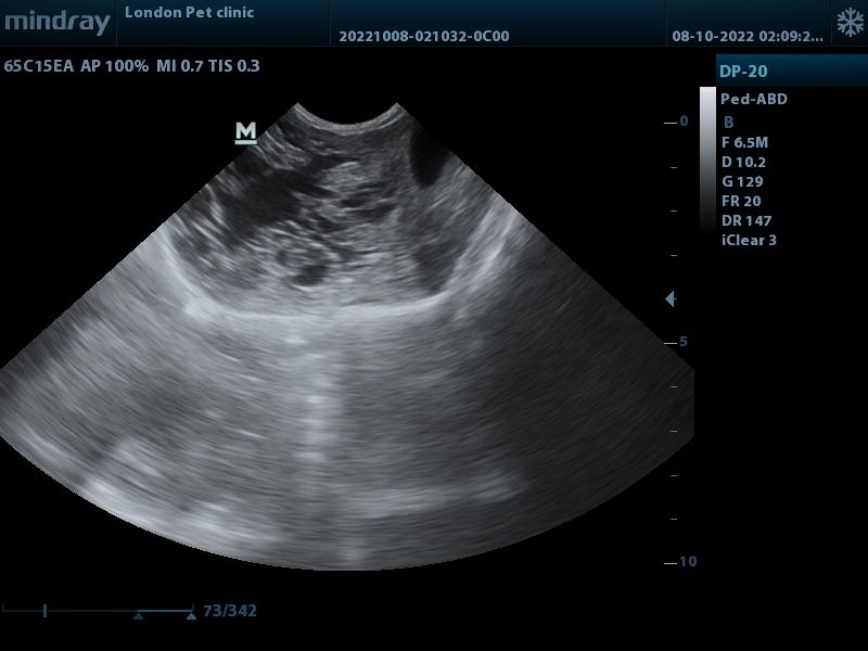
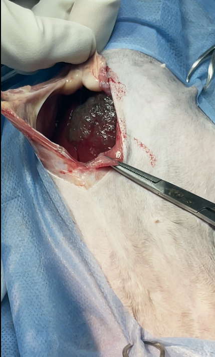
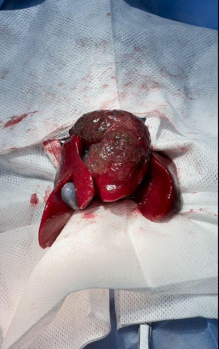
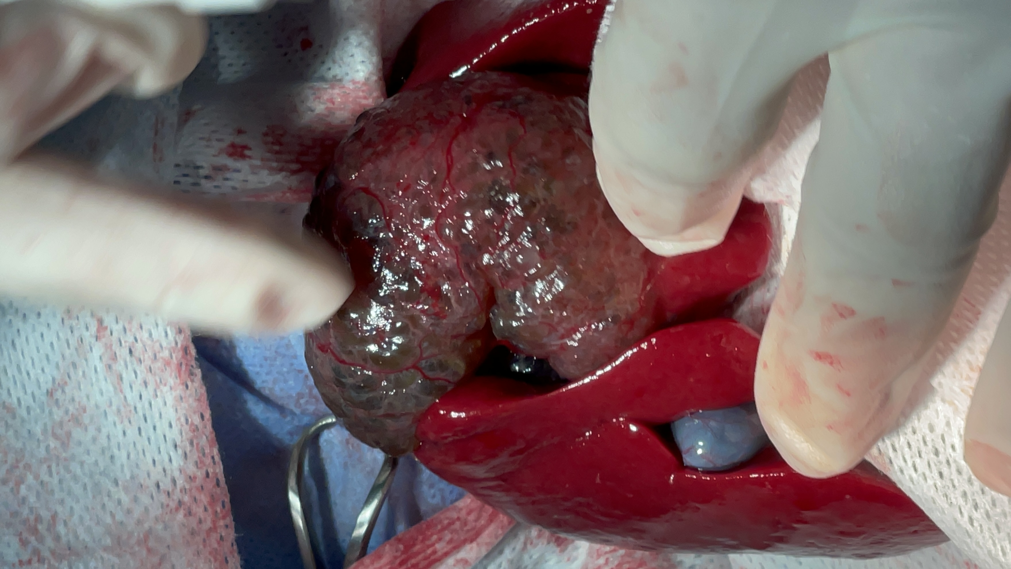
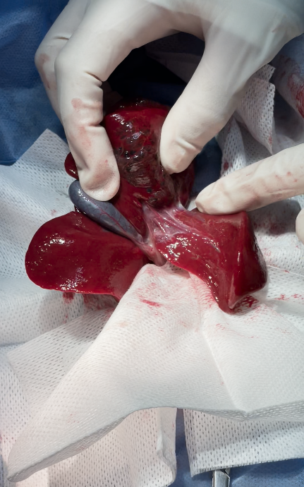
Hepatobiliary (or biliary) cystadenoma is a benign epithelial neoplasm originating from the intrahepatic or extrahepatic bile ducts. It is characterized by multiloculated cystic masses lined by biliary-type epithelium and often filled with serous or mucinous fluid

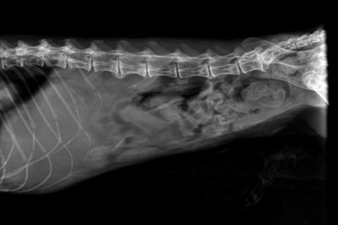
It is most often reported in middle-aged to older female cats and typically presents as a slow-growing hepatic mass

History

Clinical Signs

Diagnostics

Surgery

Histopathology

14 years old cat showing gradual onset of lethargy, reduced appetite, and mild weight loss over several weeks. The owner also reported occasional vomiting and abdominal enlargement noticed recently. There was no known history of toxin exposure, trauma, or previous hepatic disease.

The cat’s vaccination and deworming status were up to date. No previous surgeries or chronic illnesses were recorded. The owner described the cat as less active and preferring to rest more than usual.

On presentation, A-Fast Scan shows the multiloculated cystic hepatic mass with thin septations and fluid-filled cavities; walls are smooth and well-defined.

  • Clinical signs are often nonspecific and develop gradually:
  • Anorexia or decreased appetite
  • Lethargy
  • Weight loss
  • Vomiting or abdominal distension
  • Occasionally, palpable abdominal mass
  • Abdominal Radiographs: reveal soft-tissue opacity in the cranial abdomen
  • Ultrasound: Typically shows a multiloculated cystic hepatic mass with thin septations and fluid-filled cavities; walls are smooth and well-defined.

Surgical excision or partial hepatectomy is the treatment of choice.
in this case only biopsy sampling was obtained due to these reasons


owner was informed intraoperatively with :

lesion was close to major hepatic vessels & bile ducts, the risk of intraoperative bleeding or biliary leakage would be high, discouraging full lobectomy.

non-invasive, benign nature: conservative management was chosen instead of aggressive surgery

The cat’s advanced age (14 years) and possible comorbidities increased anesthesia and surgical risks

owners declined extensive surgery for risk reasons, opting for palliative management


Microscopic examination of the hepatic tissue revealed multiple cystic structures lined by a single layer of cuboidal to columnar epithelial cells resembling biliary epithelium. The cysts contained eosinophilic to lightly basophilic fluid and were separated by thin fibrous connective tissue septa.

The epithelial lining showed minimal to no cellular atypia

The surrounding hepatocytes were well differentiated, and there was no necrosis or inflammatory infiltration suggestive of malignancy.

These findings are consistent with a diagnosis of Hepatobiliary (Biliary) Cystadenoma — a benign cystic epithelial neoplasm of biliary origin

Surgery

.

.

.

.

.

.

.

.

.

.

.

.

.

.

.

.

Explore Our New uploads

RECENT SURGICAL PROCEDURES

© All Rights Reserved