C5-C6 IVDD - Cervical Ventral Slot Approach

Intervertebral Disc Disease (IVDD) is one of the most common spinal disorders in dogs, characterized by the degeneration and subsequent herniation or extrusion of the intervertebral disc material, leading to spinal cord compression and neurological dysfunction

The disease is classified into Hansen Type I and Hansen Type II disc disease, based on the nature and progression of disc degeneration


In Hansen Type I IVDD, as observed in this case, the nucleus pulposus undergoes chondroid degeneration and becomes calcified. Sudden extrusion of this degenerated material through the annulus fibrosus results in acute spinal cord compression, inflammation, and occasionally hemorrhage


History

Clinical Signs

Diagnostics

Surgery

Surgical Approach

Zeus presented to our clinic with an acute onset of severe neck pain and marked ventroflexion of the head. The patient was non-ambulatory and unable to bear weight on all four limbs, indicating complete tetraparesis.

Neurological examination revealed UMN deficits with loss of proprioception in his 4 limbs (non-ambulatory) urine retention and constipation, with normal LMN reflexes So, lesion is localized from C1-C5

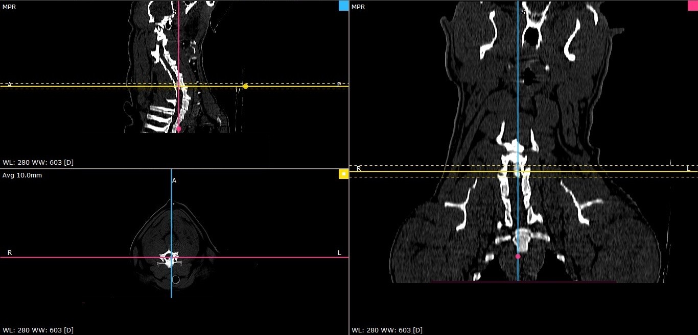
Zeus was submitted for Cervical CT scan CT revealed disc extrusion (Hansen type 1) between C5-C6 with 66% spinal compression with intra-dural hemorrhage.

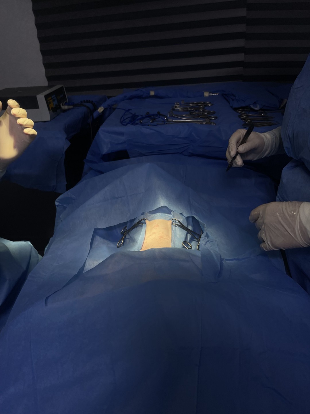
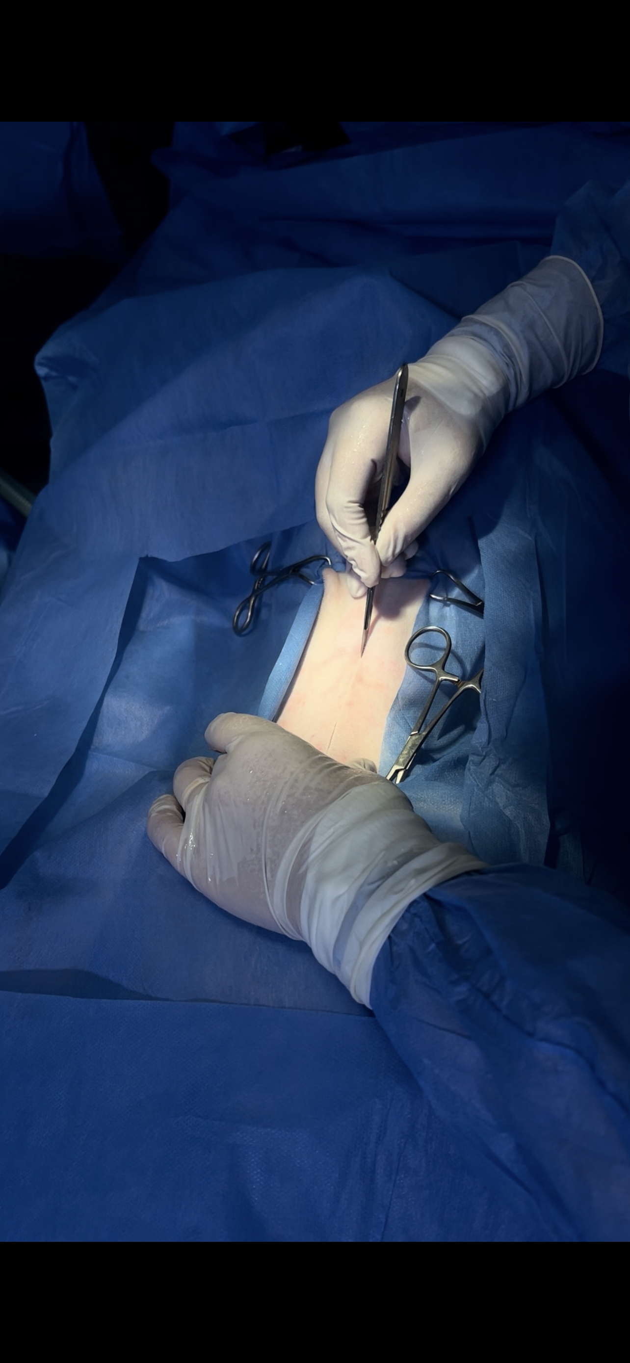
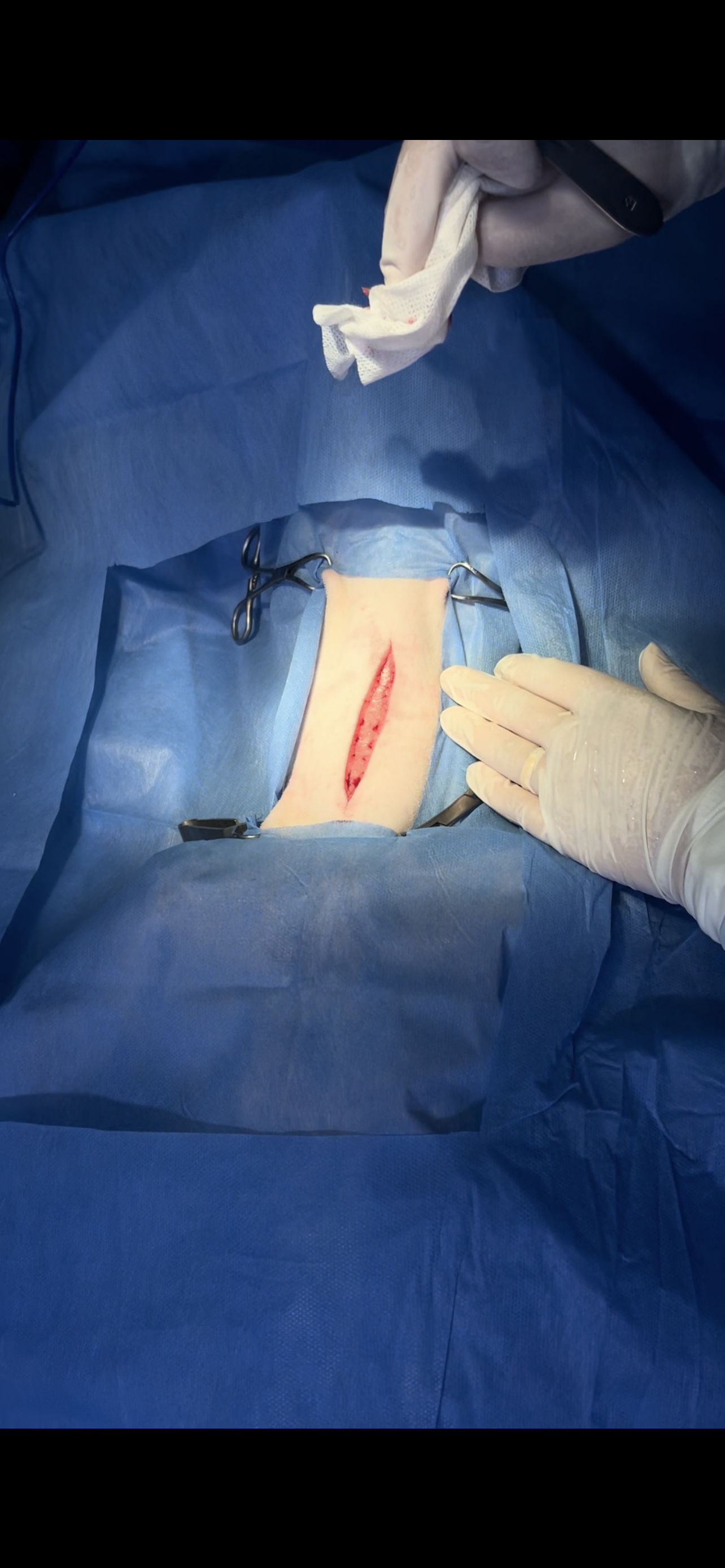
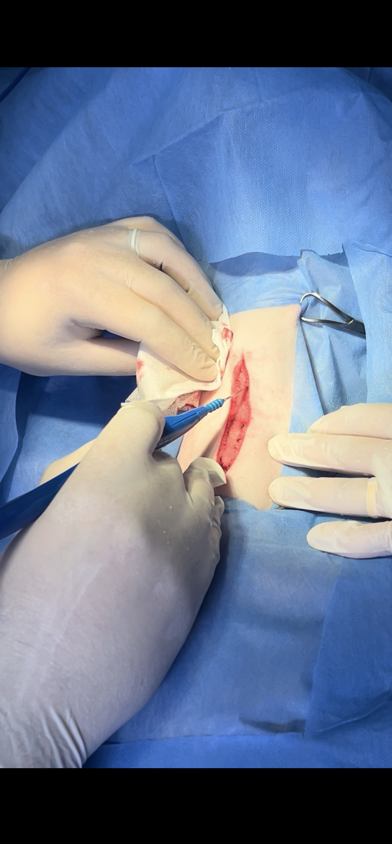
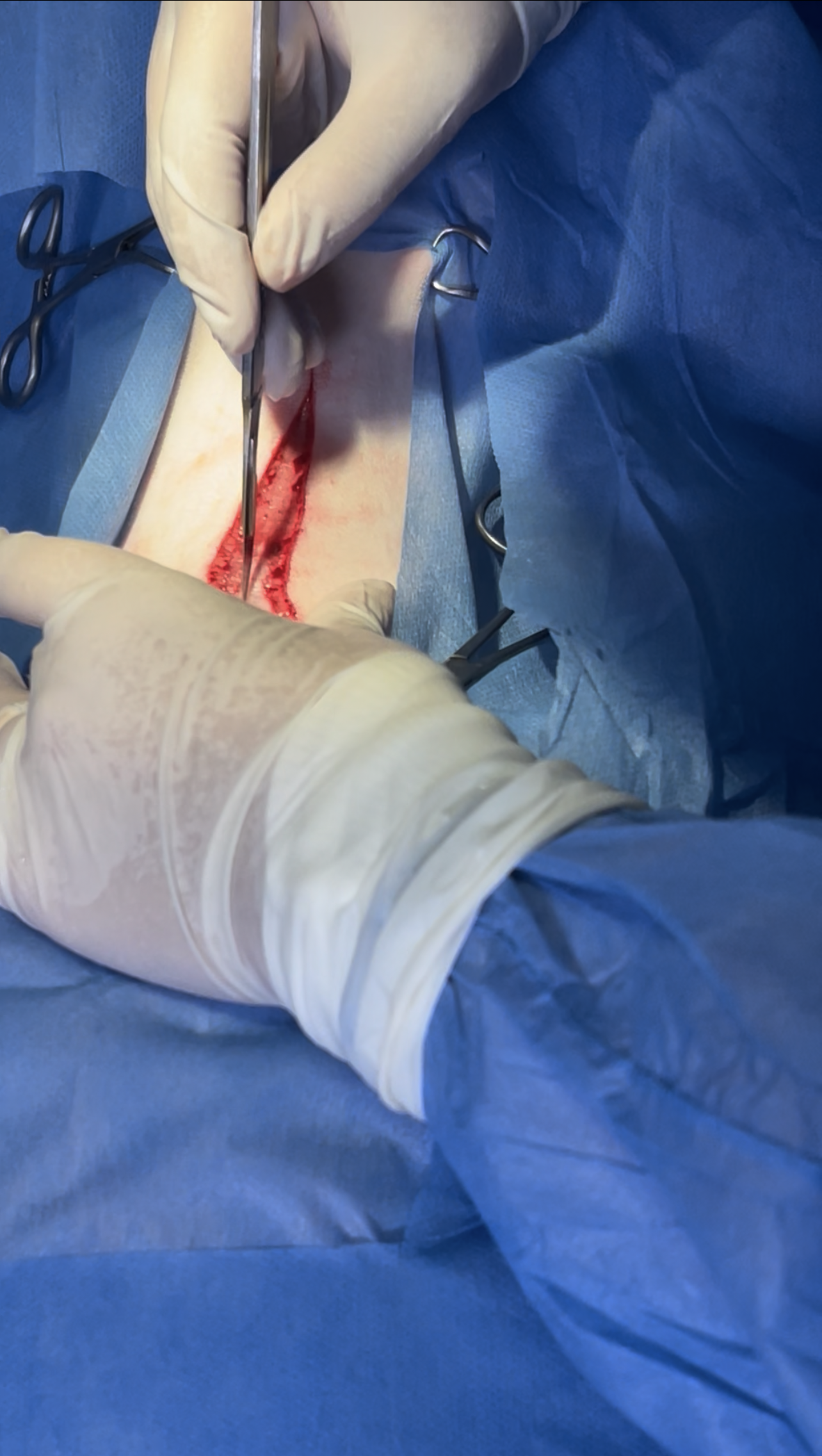
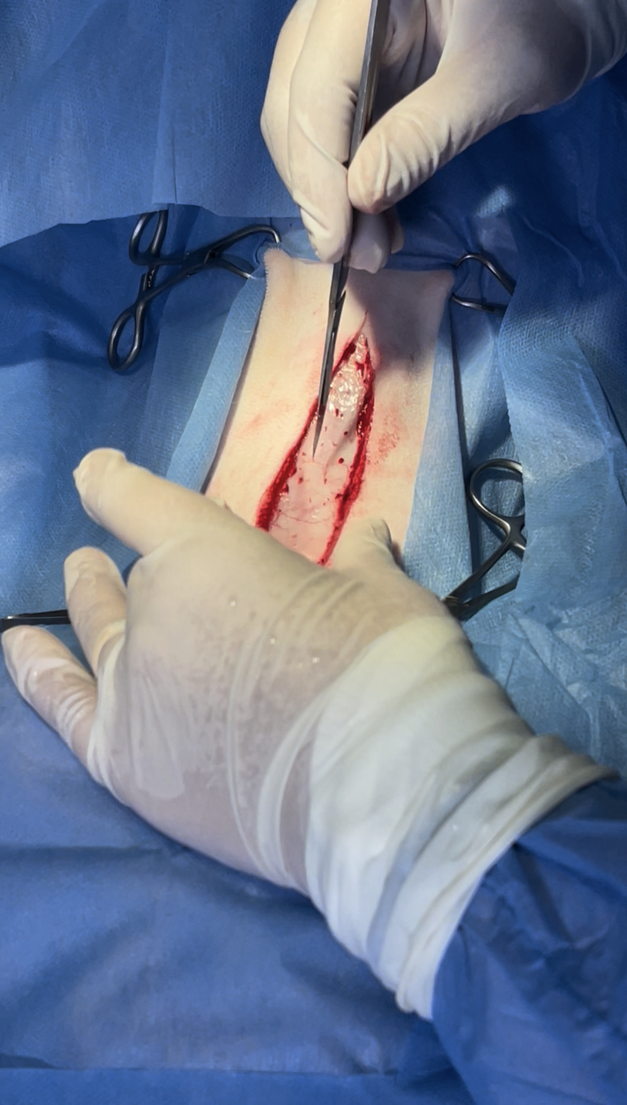
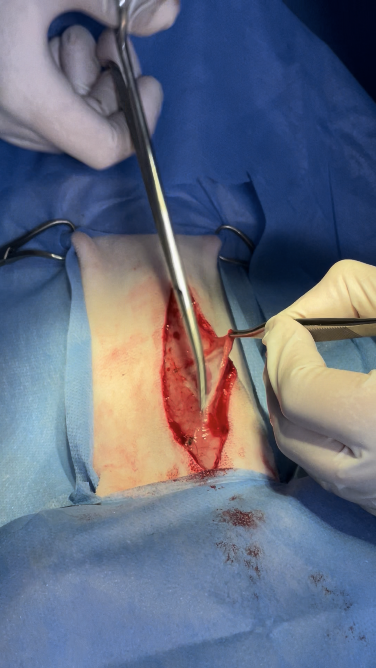
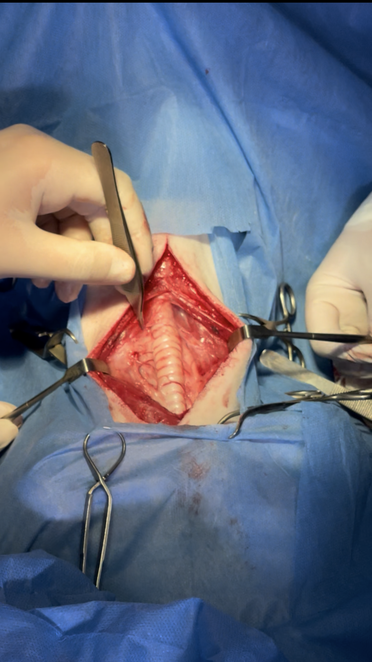
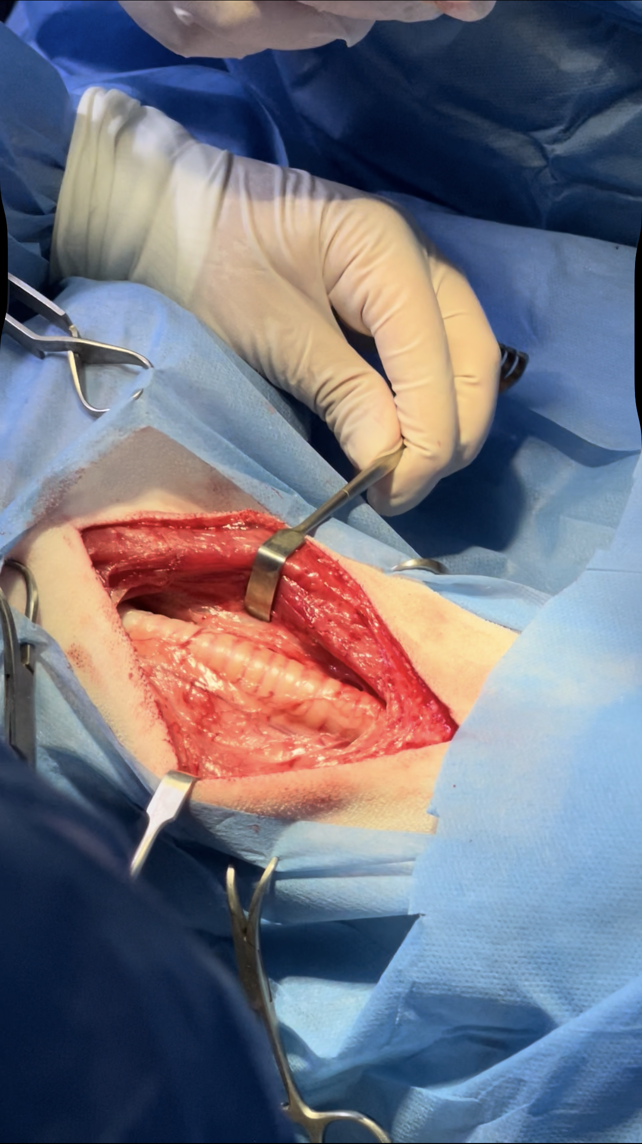
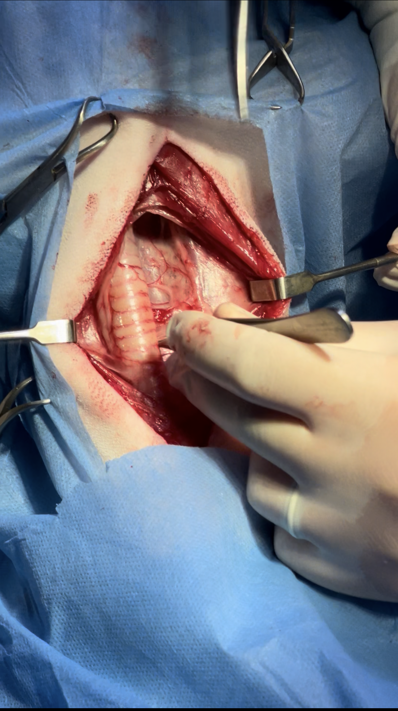
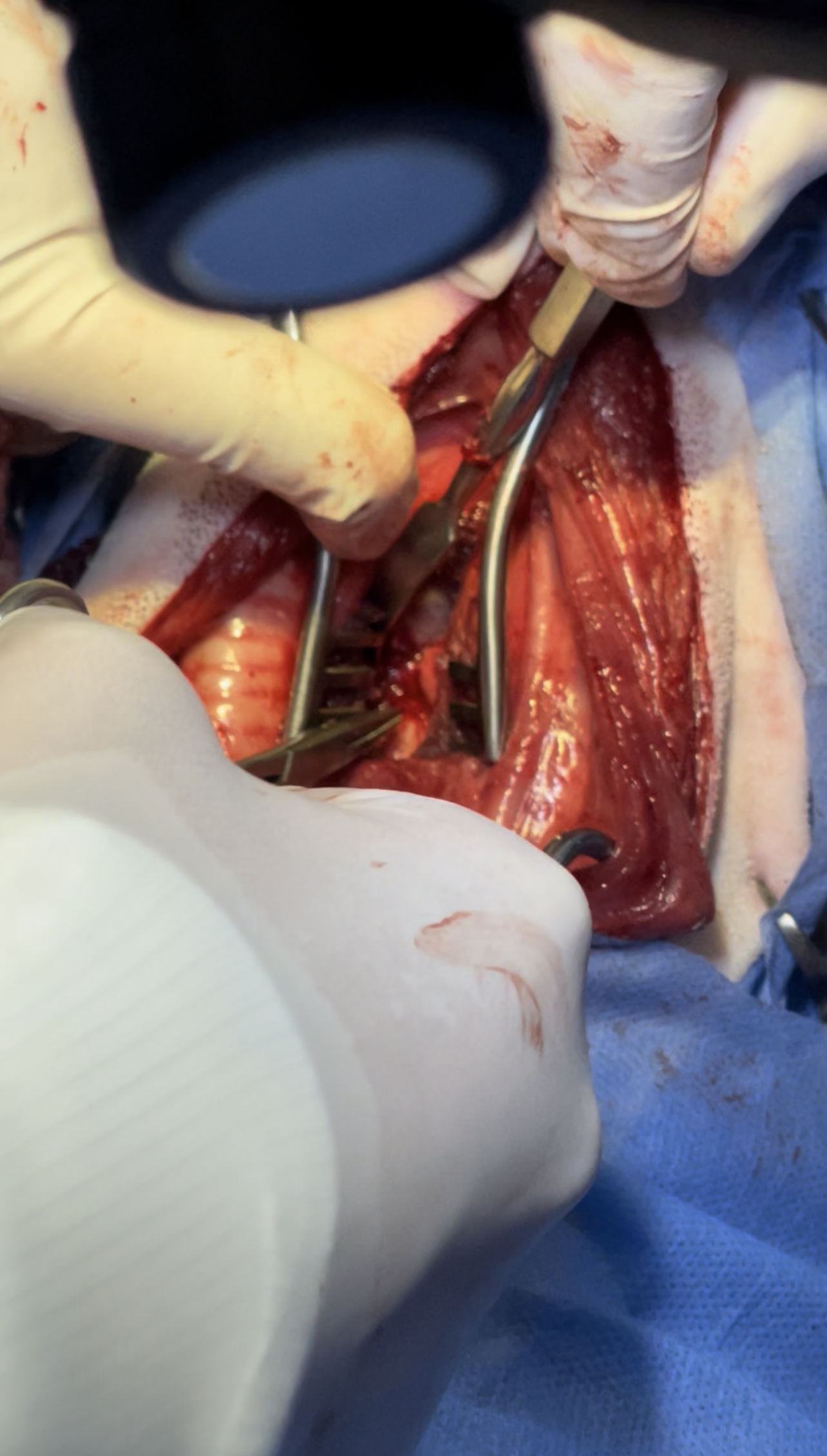
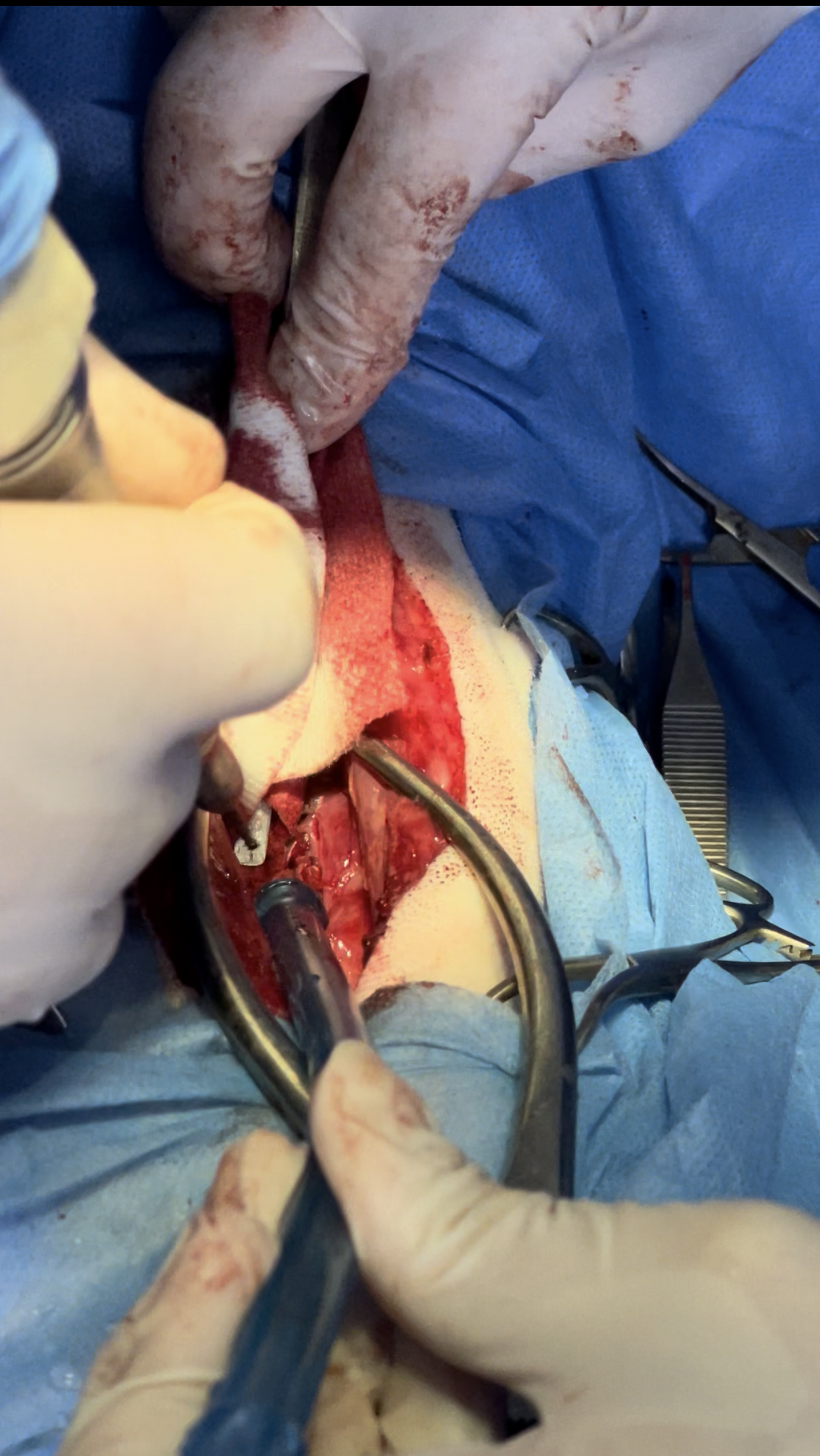
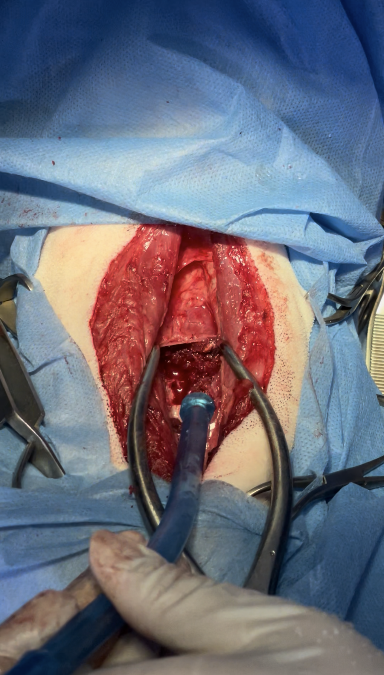
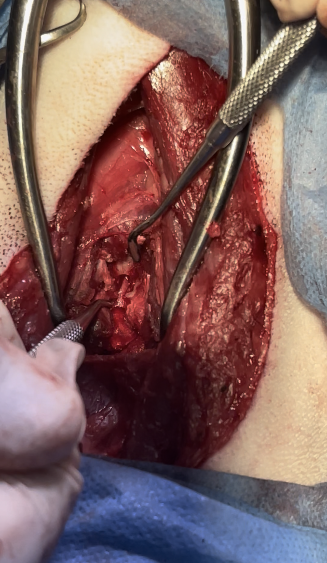
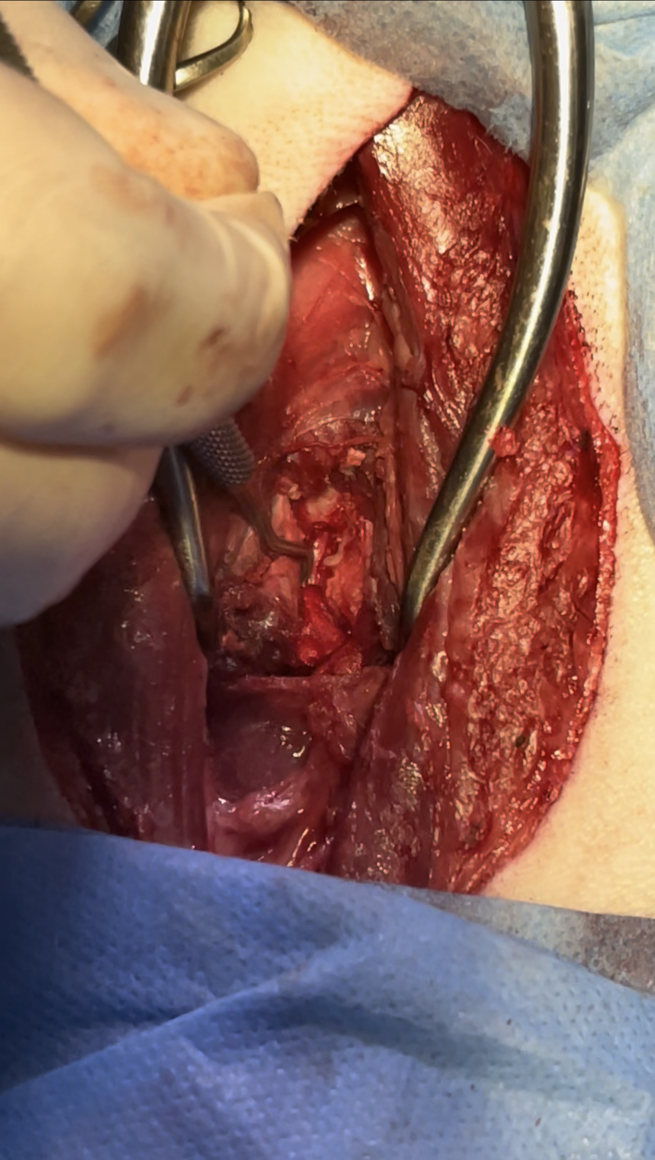
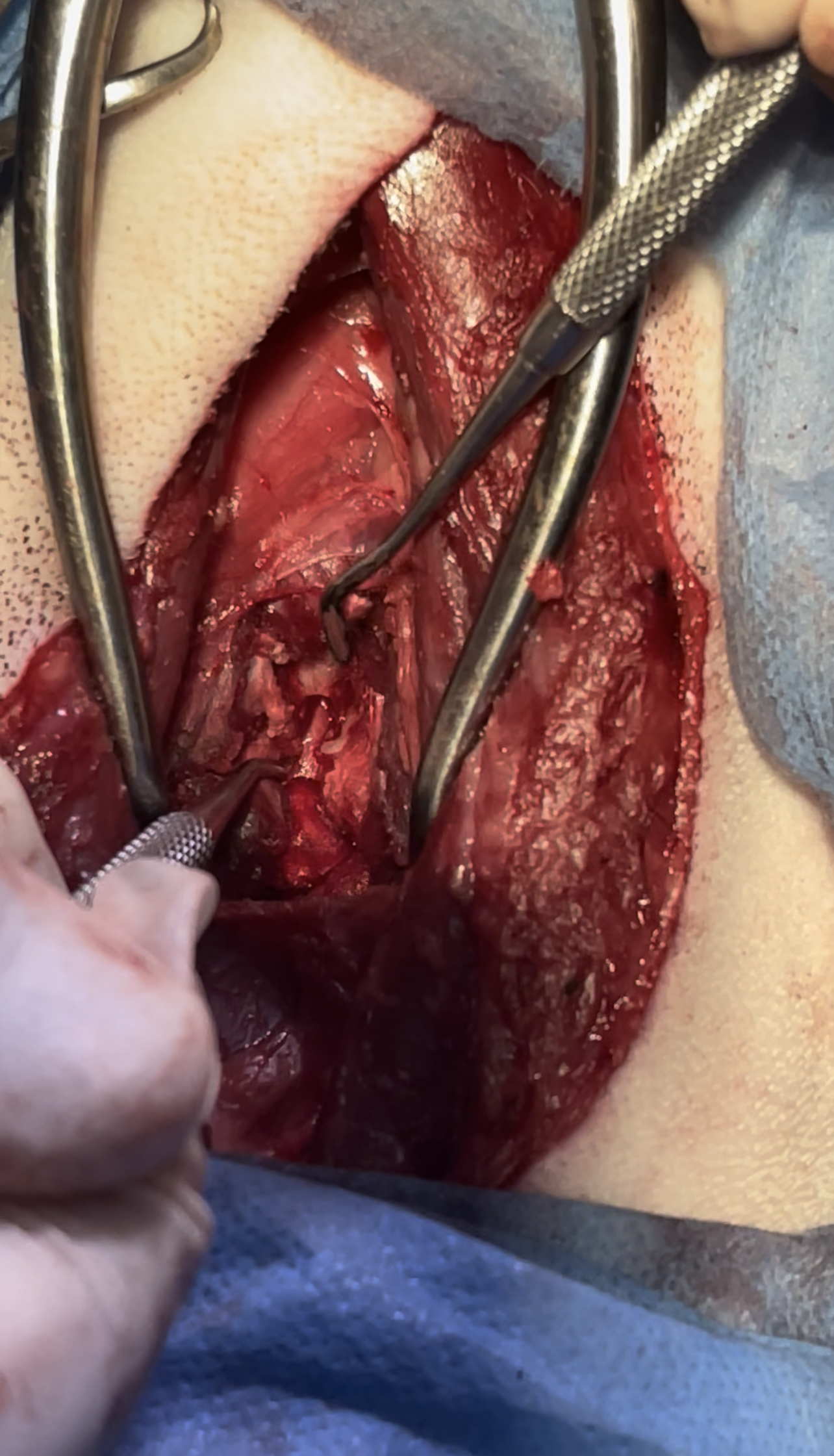
Zeus admitted to surgery Ventral slot approach was planned for removal of extruded disc material and decompression of the spinal cord surgery went successfully and he was able to take his first steps one day after the operation and regain his ability to walk after 3 days

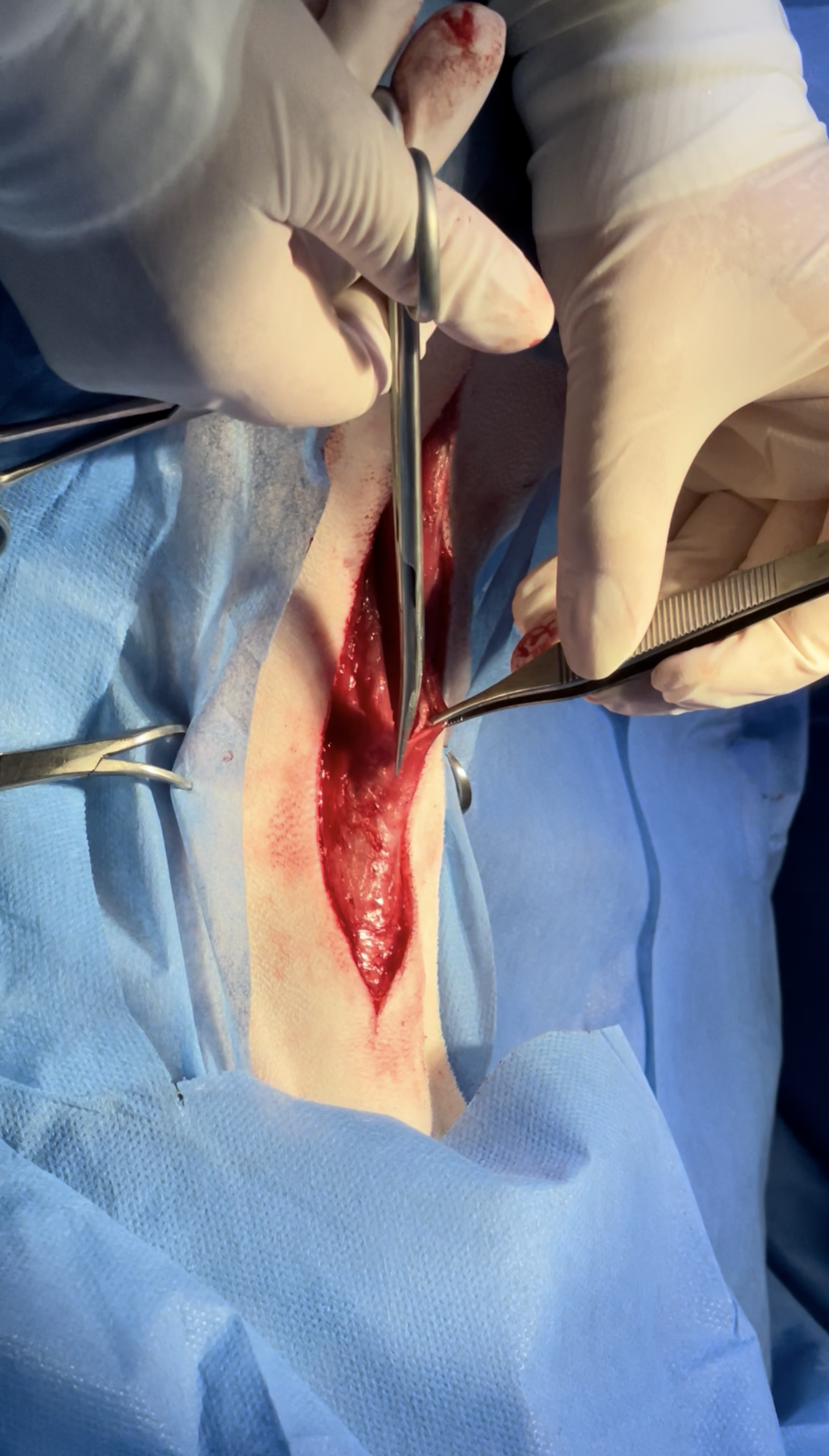
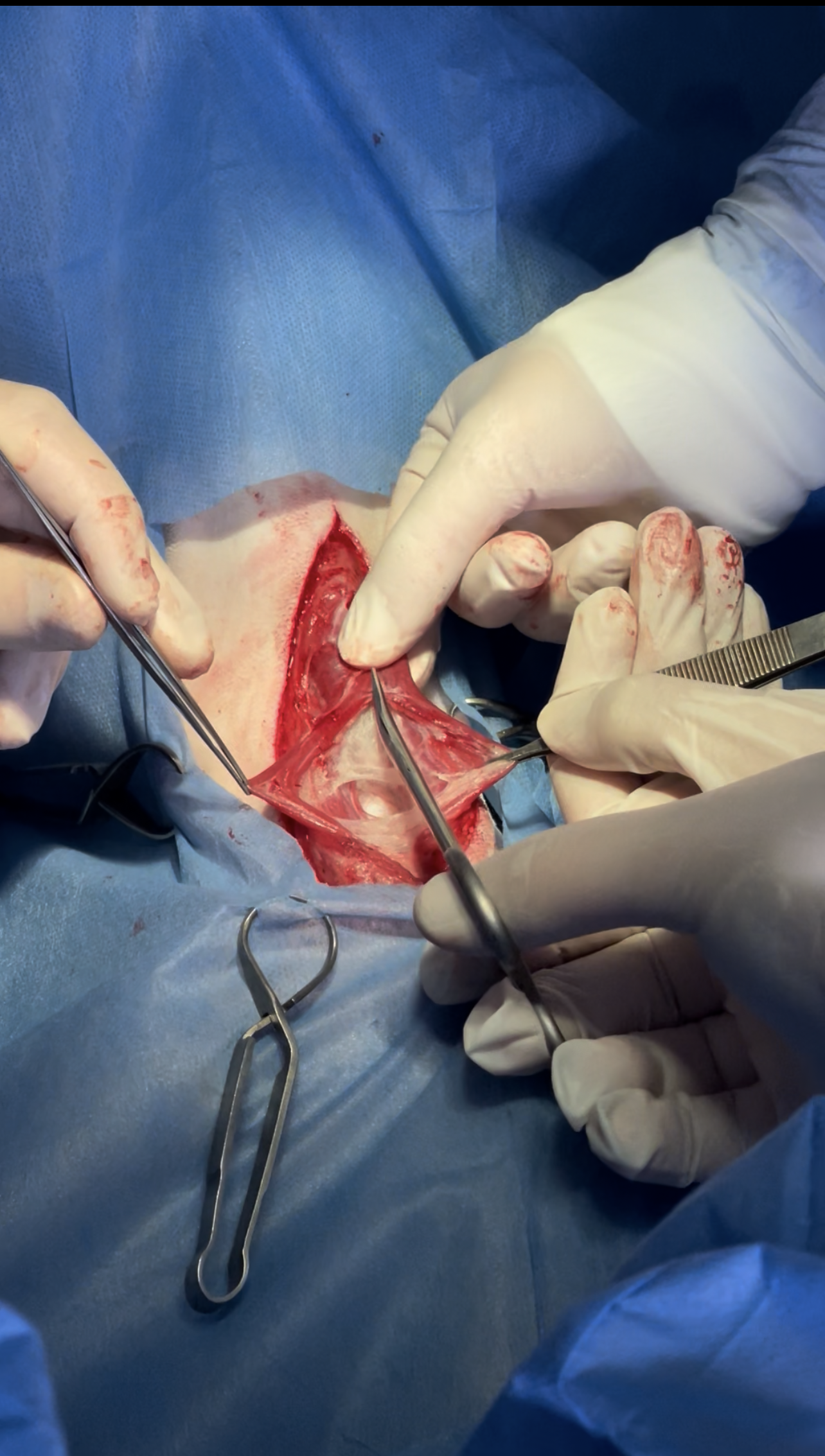
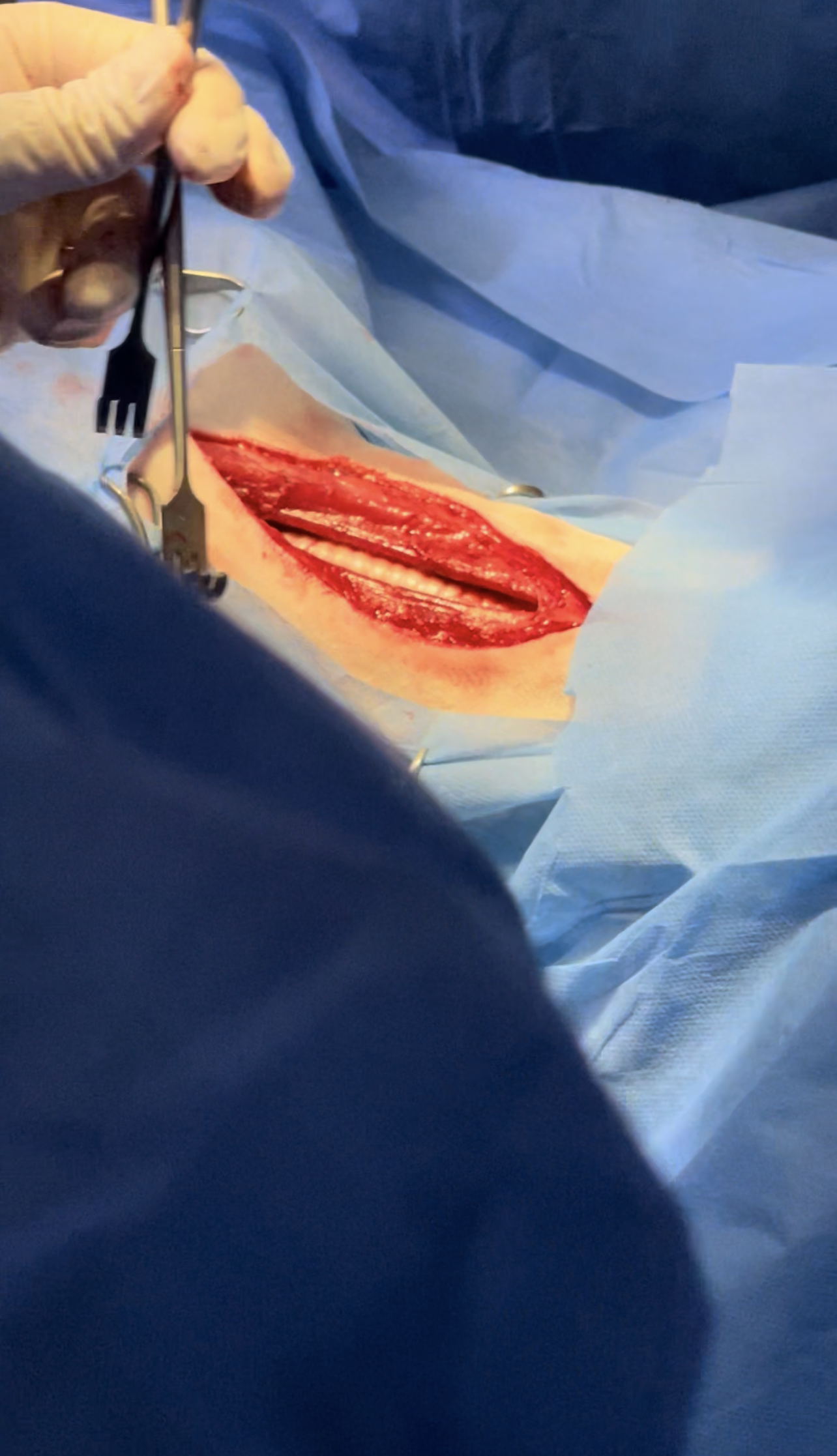
Approach to Cervical Vertebrae C2-C7 Through a Ventral slot :

  • Ventral midline incision, the incision of the skin incision extends from the manubrium to the larynx.
  • Continuing the skin incision through the subcutaneous tissues, small transverse bundles of the sphincter colli superficialis muscle are identified and transected in the ventral midline. is deepened by midline separation of the sternocephalicus muscles and the underlying sternohyoideus muscles.
  • With separation of the median raphe between the paired sternohyoideus muscles, the caudal thyroid vein is found in the fascia overlying the trachea.
  • The caudal thyroid vein should be preserved, and lateral branches of the vein arising from the adjacent right sternohyoideus muscle are divided and cauterized as necessary.
  • Lateral retraction of these muscles exposes the trachea, esophagus, deep cervical fascia, carotid sheath, and internal jugular vein.
  • Left lateral retraction of the trachea and esophagus using nontoothed retractors allows blunt dissection close to the trachea through the deep cervical fascia to the longus colli muscle, which covers the ventral surfaces of the cervical vertebrae.
  • Care should be taken not to injure the recurrent laryngeal nerve or the esophagus during this dissection.
  • The right carotid sheath containing the right carotid artery, the vagosympathetic nerve trunk, and the internal jugular vein is usually retracted to the right side of midline,
  • A short transverse incision is made through the longus colli tendon
  • Separation of longus colli muscle fibers overlying each ventral crest exposes the disk.
  • This opening into the disk may have to be enlarged for disk curettage.

Surgery

.

.

.

.

.

.

.

.

.

.

.

.

.

.

.

.

.

.

.

.

Explore Our New uploads

RECENT SURGICAL PROCEDURES

© All Rights Reserved